Ongoing · Spectrum Lab, IISc Bengaluru
Work in progressFundusFlow++
Controllable generation of retinal fundus images together with their anatomical annotations
Spectrum Lab, Indian Institute of Science (IISc), Bengaluru · Home institute: IISER Bhopal
What This Project Is About
Deep learning for retinal imaging is bottlenecked by annotation, not by images. Labelling retinal anatomy is slow, needs expertise, and the labels that matter most clinically, down to which vessels are arteries and which are veins, are the most expensive of all.
FundusFlow++ approaches this from the generative side. Rather than generating a fundus image and trying to annotate it afterwards, the system generates the anatomy and the photograph together, so every synthetic sample arrives with a pixel-perfect, clinically meaningful label attached. The image synthesis is built on flow-matching generative models[1].
My work extends an earlier system from the lab along two lines: making the generated anatomy richer and more clinically informative: in particular, distinguishing arteries from veins rather than treating all vessels alike, and making that anatomy anatomically consistent and directly controllable, so you can ask for a specific layout instead of sampling and hoping.
Controllable Anatomy
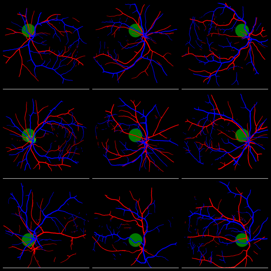
A synthetic dataset is much more useful if you can steer it rather than only sample from it. Here the optic disc is placed at nine different positions across the retina, and a complete, coherent vessel layout is produced around each requested placement.
Anatomy Taking Shape
Two samples, animated as they are synthesised.


Sample Outputs
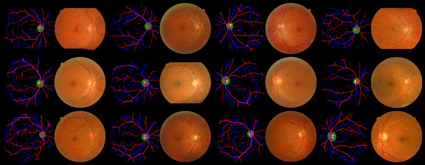
Each sample is an anatomy map paired with the fundus photograph synthesised from it, so the label and the image are produced as one unit, so the annotation is exact by construction rather than estimated after the fact.
Status
The pipeline runs end to end and produces controllable, artery/vein-labelled samples. Current directions include broader evaluation, and extending the system to pathological retinas rather than healthy ones only. A manuscript is in preparation; code and full results will follow publication.
References
- Y. Lipman, R. T. Q. Chen, H. Ben-Hamu, M. Nickel, M. Le. Flow Matching for Generative Modeling. ICLR, 2023.
- G. Lepetit-Aimon, C. Playout, M. C. Boucher, R. Duval, M. H. Brent, F. Cheriet. MAPLES-DR: MESSIDOR Anatomical and Pathological Labels for Explainable Screening of Diabetic Retinopathy. Scientific Data, 2024.
- K. Jin, X. Huang, J. Zhou, Y. Li, Y. Yan, et al. FIVES: A Fundus Image Dataset for Artificial Intelligence based Vessel Segmentation. Scientific Data, 2022.
Method references are held back until the manuscript is public.